
Spinal Stenosis and Bulging Disc?

Spinal Stenosis and Bulging Disc?

Paul K is a sprightly young 72 year old who enjoyed tennis, walking and the outdoor life. About  12/52 ago he started to feel pain in both his legs and electric shocks going down the back of both legs. Gradually he was unable to continue with outdoor activities and spoke to his general practitioner about his problems.

The GP thought he had some neurological problems based on the symptoms that he was describing and referred him for radiological examination and consultation with an orthopaedic specialist.


This is a summary of the MRI report 05 March 2024

  • normal appearances of the visualised distal cord down to the conus at the L-1 level. Degenerative changes in the marrow signal.
  • Mild degenerative changes down to L2/L3
  • L3/L4 – there is an extruded disc which causes moderate to severe central canal with almost complete effacement of the CSF within the thecal sac at this level. Mild bilateral neural foraminal stenosis.
  • L4/L5 – minor degenerative disc bulge pressures the right L5 nerve in the right lateral recess and contacts the left L5 nerve. There is also severe right neural foraminal stenosis with potential right L4 neural compromise.
  • L5/S1 – minor degenerative disc bulge which contacts the L5 nerves in the far lateral position bilaterally.
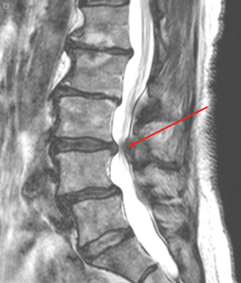
    Conclusion:

    • There is moderate to severe central canal stenosis at the L3/L4 level.

    • The patient’s bilateral posterio-lateral leg pain could be caused by the potential bilateral L5 compromise.

    Paul telephoned me from the UK concerned about his symptoms and his diagnosis from the radiologist. He asked my advice on whether I was able to help him instead of having surgery as the orthopaedist suggested.

    He reported that there was no intermittent claudication associated with spinal stenosis, that both his legs felt strong and that he only experienced an electric shock in his legs and feet when he ran or was going downstairs. He experienced low back pain when standing for long periods.

    I suggested that the symptoms did not match the radiologist comments and maybe the MRI pathology was incidental to his current symptom profile. He decided to fly to Portugal for a consultation and physical examination.

    On examination I found he had pelvic instability, a physiological short-leg, a functional scoliosis of the lower spine, trigger points in the left sacroiliac ligament and muscle spasm running from the left sacroiliac joint up to the left occipito-atlantal joint just below the skull.

    Treatment involved prolotherapy injection to the left sacro-iliac ligament.  After three minutes the low back pain had disappeared, the spinal muscle spasm had gone and the legs were level. Three days later I did osteopathic manipulation to restore functional integrity of the spine. When he left, after seven days, he reported all his symptoms had disappeared apart from a mild low back pain which has been with him for many years.

    In conclusion:

    If Paul had followed the orthopaedist’s advice and had surgery for the spinal stenosis and the disc protrusions it might have been considered “failed back surgery” because the operation would have treated the results and not the cause.

    It is important for patients to seek advice from a practitioner working and practising a different medical paradigm before consenting to surgical mutilation and statistical profiling.

    We, at the Centre for Bioregulatory Medicine, considers everyone is an individual and decides the most appropriate treatment according to the patient’s needs. Surgery is sometimes the only solution if the pathology is severe but unnecessary surgery is a crime.

    Criminal law does have a role to play when surgeons abuse trust and harm patients, but understanding when non-fatal surgical harm should be treated as a criminal matter is a complex issue.


    Cardiac arrhythmia and panic attacks

    Cardiac arrhythmia and panic attacks

    Is it arrhythmia or panic attack (the chicken or the egg story)?

    Diane H, a 54 year old business woman, attended the clinic complaining of a racing heart, dizziness, difficulty in breathing, tingling in the hands, “butterflies” in the stomach and hot flushes. At the time she attended the clinic she was free of symptoms but they were occurring on a regular basis which made her feel there was something seriously wrong with the heart or head.

    When she visited the doctor, again, the symptoms disappeared and even though he attached her to a heart monitor and took her blood pressure there was nothing significant to pass comment.

    Her job was quite stressful and it was this that the doctor thought triggered the panic attacks and the associated cardiac arrhythmia.

    Diane was prescribed amitriptyline by her doctor to help with the panic attacks as he considered her problem was more psychological than cardiac.

    The frequency of the panic attacks increased whilst taking the drugs and therefore came to see me for a second opinion.

    Comment:

    Her symptoms are consistent with compromise of the vagal nerve. This nerve leaves the base of the skull between the carotid artery and jugular vein and travels virtually the whole length of the body sending nerve signals to the heart, lungs, abdomen and digestive tract.

    On examination Diane displayed left sacroiliac ligament laxity, pelvic instability, leg length inequality, muscle spasm running from the left sacroiliac joint to the base of the skull and muscle spasm in the left buttock.

    L

    sacro-iliac joint dysfunction

    L

    muscle spasm running the whole length of the spine

    L

    twitch response on palpation

    L

    neural compromise to the hands

    Please click on icon to show label

    The ligamentous instability of the left sacroiliac joint caused a compensatory muscle spasm in the left buttock and the spine. This compromised the vagus nerve which controls the heart, the lungs and the gastrointestinal tract. The functional scoliosis caused by the leg length difference compromised the neck and the nerves that supply the hands.

    Abdominal Distension and Flatulence

    Abdominal Distension and Flatulence

    Maria S, 34 year old mother of two, attended the clinic complaining of abdominal pain, distension and gas. She reports that this problem started shortly after the birth of her second child nine months ago.

    She changed her diet in the hope that this would help with the bothersome gas and distension. The feeling of fullness and pressure in the abdomen continued with an observable increase in the size of the abdomen.

    She has undergone various medical tests to ascertain whether there is pathology, viral or bacterial infection, allergies and food intolerances.

    All gave negative results.

    On examination:

    Pelvic imbalance with twitch response in the right sacro-iliac ligament, pain radiating into right groin, muscle spasm in right ilio-psoas muscle and tenderness on palpating the right lower abdominal quadrant.

    My conclusions:

    The pelvic imbalance, possibly as a result of the birthing process of the second child, was maintaining a muscle spasm in the right iliopsoas muscle which was mechanically obstructing the passage of faecal matter and gases through the colon.

    Treatment:

    Prolotherapy injection was given to the right sacro-iliac ligament. After three minutes most of the gas has dissipated and the patient felt more comfortable. Over the next five days the patient reports that the abdominal distension had gone and that she had normal bowel movement. Osteopathic manipulation was given seven days after the prolotherapy treatment to realign the spine and pelvis.

    Disclaimer:

    Many of the conditions presented within this prolotherapy section of functional disorders is where, in my opinion as an osteopathic physician and prolotherapist, I believe that the structural imbalance presents itself as a disturbance in function. Therefore, after the patient exhausts all the medical tests, I use physical medicine as a choice of therapy.

    .

    Damaged right lateral knee ligament

    Damaged right lateral knee ligament

    Lorem Ipsum is simply dummy text of the printing and typesetting industry. Lorem Ipsum has been the industry’s standard dummy text ever since the 1500s, when an unknown printer took a galley of type and scrambled it to make a type specimen book. It has survived not only five centuries, but also the leap into electronic typesetting, remaining essentially unchanged. It was popularised in the 1960s with the release of Letraset sheets containing Lorem Ipsum passages, and more recently with desktop publishing software like Aldus PageMaker including versions of Lorem Ipsum.

    Period Pains (uterine latero-flexion)

    Period Pains (uterine latero-flexion)

    Vivian, a female of 19 years of age, came to the clinic complaining of regular but painful periods that has gradually become worse over the last 10 years

    She has visited her gynaecologist many occasions who prescribed anti-inflammatory drugs and the contraceptive pill. Within two periods she started to feel nauseous and suffered from migraines which, I feel, is a side effect of the drugs she was prescribed.

    Approximately three months ago she was sent for an MRI of the pelvis to exclude pathology and to try and understand why she was having such painful periods.

    Information clinical: Intense, disabling dysmenorrhea.

    EXAMINATION REPORT

    • Bladder is very full, thin-walled and regular
    • Uterus in retroversion, anteflexion and slight right latero-shift, measuring approximately 8 x 4 x 3.5 cm in longitudinal, transverse and anteroposterior axes respectively.
    • The myometrium is homogeneous. There is no anomalous thickening of the junctional zone and there are no nodular lesions attributable to leiomyomas. The endometrium is regular, with an estimated normal thickness of 4 mm.
    • A small retention cyst measuring 5 mm was identified in the endocervical canal.
    • Ovaries in their usual topography, with preserved morphostructure, with some infracentimeter follicular imageries visible bilaterally.
    • No iliac or inguinal adenomegaly was identified.
    • Free fluid in the fundus of the Douglas bag, considered physiological.

    Please click on icon to show label

    From the radiologist report there is an indication that the uterus is not in the correct position. We can see  uterus is sidebent (latero-version) to the right producing congestion around the right ovary. The utero-sacral ligament is hyperintensive indicating tissue changes.

    \

    Utero-sacral Ligament

    \

    Sacrum

    \

    Bladder

    \

    Right latero-verted Uterus

    \

    Ovarian congestion

    \

    Rectum

    L

    Bladder

    L

    Uterus

    L

    Cyst

    L

    Vagina

    L

    Sacrum

    L

    Pubis

    Please click on icon to show label

    From the lateral view we can see a small cyst at the cervico-vagina junction along with anterio-flexion of the uterus and flexion of the coccyx.

    The bladder is full and enlarged obstructing the uterus and surrounding structures.

    Comments:

    According to Dr Andrew Taylor Still, the founder of osteopathic medicine, structure (anatomy) affects function (physiology). The MRIs of Vivian’s pelvis shows structural anomalies which will interfere with the normal function of uterus and ovaries. The cyst may also prevent free flow of the menses. The utero-sacral ligament attaches to the sacro-spinous ligament which demonstrated a twitch response when palpated.

    The treatment involved prolotherapy to the attachments of the sacro-spinous ligament followed a week later by osteopathic manipulation to the sacrum and coccyx.

    It took a further two periods before Vivian felt pain relief during her periods. Osteopathic manipulation to the spine and pelvis continued during this period of time. Another MRI is recommended in six months time to check position and function of the uterus.

    .